The role of neutrophils in wound healing and elasticity of the skin of wild-type and TgfbrN mice studied by atomic force microscopy
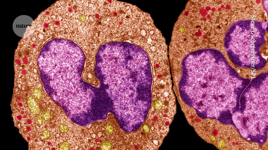
Scientists have known for a long time that the neutrophils cause chemical warfare by releasing toxins. But the new work, published today in Nature, reveals “an additional role for neutrophils here that we had not appreciated”, says Niki Moutsopoulos, a clinical immunologist at the National Institutes of Health (NIH) in Bethesda, Maryland, who was not involved with the study. She says that the results show that neutrophils help in wound healing.
Neutrophils are known to poison invading microbes and to swallow them. But these strategies cause “collateral damage” to the body by killing healthy bystander cells, Hidalgo says.
a, Representative images of neutrophils (green) stained for Col3a1 (red) in the skin of WT control (CreNEG Tgfbrflox littermates, 5 images from 2 mice) and TgfbrΔN mice (9 images from 3 mice), which we used to quantify the number of neutrophils per volume area is shown at right. Mean values are presented as a comparison of two-tailed Student’s t-test. The skin of wild-type control and TgfbrN mice was characterized through hematoxylin-eosin staining. The images are quantified in the graphs. P-values were determined by a two-sided unpaired Student’s t-test. The examples of fibre reconstruction are shown in the attached table, as well as the distribution of fiber width in the histogram below and in the Extended Data Fig. Control versus iDTR mice. The two-sided t-tests were used to make the P-values. The structures of the atomic force microscope set-up are used to measure the stiffness of tissue samples and the elasticity of Young’s modulus in lung, intestine and skin. The Young’s modulus value is calculated from individual force–distance analysed curves per mouse. Two-sided unpaired Student compared the data. The height images are related to Young’s modulus maps of skin from the control and TgfbrN littermates. There were images of the ear skin, which showed a variety of factors in the subepidermal and lower dermis regions. There are yellow circles around the sub epidermal region. Data are from 2 independent experiments and pertain to the quantification of fibre size in the skin of mice. P-values determined by Kruskal-Wallis non-parametric test. g, Per cent of ‘large’ fibres in the subepidermis and lower dermis from the images in f. Data are presented as mean values ± s.e.m., and p-values were determined by Kruskal-Wallis non-parametric test. h, Permeability assays in control (n = 7) and TgfbrΔN mice (n = 7) measured by FITC–dextran injected either intratracheally (for lung), or by oral gavage (for gut) in CreNEG control (n = 8) and TgfbrΔN mice (n = 8). Evans blue given intravenously to control (n = 11) and TgfbrΔN mice (n = 5) and then measured in the indicated tissues. All the controls here had Tgfbrflox littermates. i,j, Stiffness (i) and permeability assay (j) using Evans blue in the ear skin of Mrp8Cre; Tgfbr2flox and Ly6GCre; Tgfbr2flox mice, as well as CreNEG; Tgfbr2flox littermates, or mice treated with anti-Ly6G to deplete neutrophils or isotype control antibody. The numbers of replicates are displayed in the figure. Two-sided Student tests comparing each method with their controls or one-way ANOVA, with a number of comparison panels on the right. Box plots in (d,h–j) show median ± interquartile; whiskers show the range from minimum to maximum. The whiskers are defined with percentiles and IQR, which are the points outside the interquartile range.